Welcome once again to the concluding part of our research work. In the previous part shared about a few days ago, we looked at some of the attributes of Nigella sativa and how they mitigate strongly against oxidative stress in humans through their antioxidant potentials. We also extended the discussion by explaining the basic things about oxidants, oxidative stress, antioxidants, and cadmium and how their presence in the body can great impact on our general well-being as humans.
Today, we will critically discuss the results of our research work and fully demonstrate how Nigella sativa (black cumin seed) offers great help in helping the body recover from tissue injuries that could be caused by various factors. Our current research work used Cadmium ( a well-known tissue toxin) to induce injury in rat models. Our interest was focused on seeing how well the body will heal and recover from the induced tissue injury with the help of Nigella sativa.
Normally, during cell or tissue injury, some common biomarkers are usually elevated. Furthermore, the injury can as well be demonstrated and seen by doing a histopathological examination of the tissue to observe the morphological changes that have occurred in the tissues. While histological changes are seen through observation under the microscope, biomarkers are mainly analyzed biochemically using various analytical procedures.
Interestingly, the study showed that Nigella sativa was able to reverse the tissue injury in some of the affected organs. The heart which was the most affected by this injury was also seeing to fully recover. All these and more are what we will be demonstrating in our discussion for today. Let's briefly look at some of the key important areas of the research work.
Research methodology
Preparation of Methanolic extracts of Nigella sativa (MENS) Nigella sativa seeds were dried and shaded from sunlight, then powdered with a grinder. Extraction was done using Babaei et al. methods with minor modifications. Five hundred grams (500g) of N.sativa powder was macerated with 2 liters of absolute (100%) methanol (as methanolic extract) for seventy-two (72) hours. The mixture was stirred in an Erlenmeyer flask for twenty-four (24) hours using a laboratory shaker.
At the end of the extraction, the extract was filtered through a Whatman filter (Whatman Clifton, NJ, USA). Finally, using a water bath set at 30°C, the solvent evaporated, and 4g of dried methanolic extracts were obtained. This was reconstituted in distilled water, used to prepare the required concentration, and stored at 4°C until when needed for use.
We further did an Acute toxicity test (LD50) to determine the median lethal dose of the extract. The results are as seen below.
Table 1: The median lethal dose (LD50) of methanolic extracts of Nigella sativa (MENS)
 Two doses, 1200mg/kg, and 5000mgkg were used to calculate the LD50 of the plant extract.
LD50 = √ A x B
A = Maximum dose with 0% mortality (1200mg/kg)
B = Minimum dosed with 100% mortality (5000mg/kg)
LD50 of methanolic extract of N.sativa seed = √1200 X 5000 = 2449.48mg/kg
LD50 of methanolic extract of N.sativa seed ≈ 2400mg/kg
The above clearly shows that the Median lethal dose (LD50) value of the extract was 2400 mg/kg which indicates that MENS is safe and is not toxic to mice, at the doses used in the experiment.
Experimental Design and procedures The twenty-five (25) male rats were grouped into (A-E) and received the following treatments daily and within 2 hours. The animals were kept under observation for 14 days before the onset of the experiment for acclimatization. The experimental protocol was approved by the institution's animal ethics committee of the University of Nigeria Teaching Hospital (UNTH/CSA. 452/VOL. 19).
Sub-acute cardiac, kidney, and liver injuries were further induced in each animal by intraperitoneal injection with cadmium chloride solution (5mg/kg), daily for 14 days.
Group A: (normal Control): No treatment was administered to this group.
Group B: (Negative Control): received CdCl2 (5mg/kg, intraperitoneal) only for 14 days.
Group C: received CdCl2 and a low dose (300mg/kg, oral) of methanolic extract of N. sativa MENS for 14 days.
Group D: received CdCl2 and high dose (600mg/kg, oral) of methanolic extract of N. sativa (MENS) (600mg/kg, oral) for 14 days.
Group E (Positive control): received CdCl2 and Vitamin C (200mg/kg, oral) for 14 days.
After 14 days, the animals were sacrificed via cervical dislocation under chloroform anesthesia. The heart, kidney, and liver were harvested for histopathological analysis. The Histopathological analysis of a tissue sample gives a better visual representation of what is happening to the tissue than does biochemical analysis. However, biochemical analysis can still supplement histopathological analysis.
Our focus was on three major organs - The heart, kidney, and liver. The excised heart, kidney, and liver tissues were processed using the paraffin wax embedding technique, sectioned at 5 microns, and stained using the Haematoxylin and Eosin [H and E] staining procedure. The histological sections were examined using an Olympus TM light microscope.
Results and findings
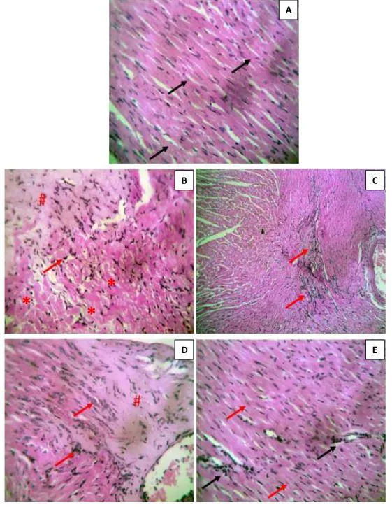
In Figure 1 as shown below, the heart of normal control rats appeared functionally and structurally normal. The cardiac fibers showed a well-conserved morphology (1A). The heart of the CdCl2-treated group (negative control) showed abnormal changes; there was evidence of fibrosis and mild infiltration by inflammatory cells. Fibers appear wavy showing signs of significant degeneration (1B).
 Figure 1: Photomicrograph of heart section. (A) Cardiac fibers (black arrows) appear normal with no degenerative changes. (B) Evidence of fibrosis (#) and mild infiltration by inflammatory cells (arrows). Fibers appear significantly wavy and damaged (*). (C) A section of the cardiac fibers appears necrotic and inflamed with infiltration by inflammatory cells (arrows). (D) Evidence of fibrosis (#) and mild infiltration by inflammatory cells (arrows). (E) Cardiac fibers (red arrow) appear normal with very mild infiltration by inflammatory cells (black arrows) [Stain: H and E; ×400].
However, the cardiac fibers of test group rats (low dose MENS at 300mg/kg) appeared normal with very mild infiltration by inflammatory cells (1C). While in the other test group of rats (high dose MENS at 600mg/kg), the myocardial fibers appear wavy; some fibers are necrotic, with the presence of leucocyte infiltration (1D). Furthermore, a photomicrograph of the heart section from CdCl2 + Vitamin C (200mg/kg), showed a normal appearance of cardiac fibers (1E).
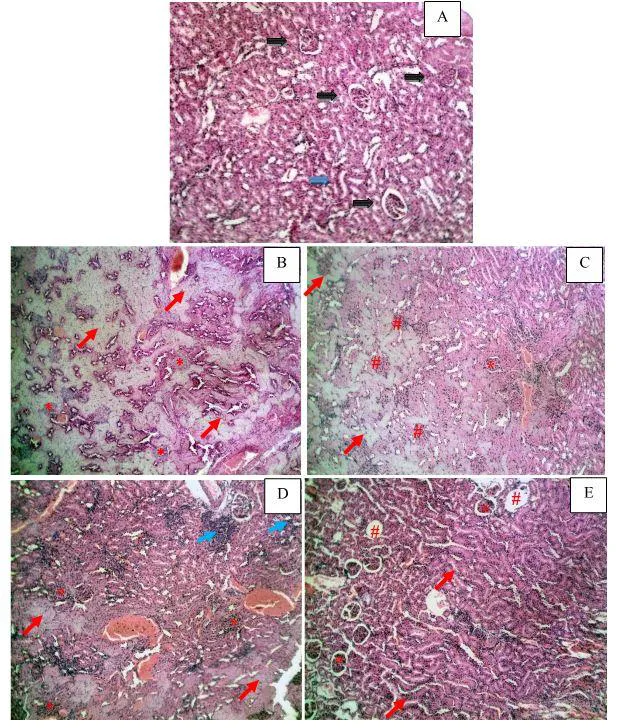
In Figure 2 as shown below, the kidney of normal control rats appeared functionally and structurally normal. The glomeruli and tubule showed a well-conserved morphology (1A). The kidney of the CdCl2-treated group (negative control) showed abnormal changes; there was severe tubular degeneration; the glomeruli are all enlarged (1B).
 Figure 2: Photomicrograph of kidney section. (A) There is a normal appearance of glomeruli (black arrows) and renal tubule (blue arrows). (B) There is severe tubular degeneration (red arrows); the glomeruli are all enlarged (). (C) Most glomeruli are enlarged and hypercellular (#); few are still normal (). (D) The tubules show signs of autolytic degeneration (red arrows); the glomeruli are enlarged () and there is infiltration by inflammatory cells (blue arrows). (E) Most tubules appear normal (red arrows); glomeruli also appear normal () with a few eroded Bowman’s capsules (#). [Stain: H and E; ×100]
However, the kidney tubules of test group rats (low dose MENS at 300mg/kg) showed mild signs of autolytic degeneration and erosions; most glomeruli were normal while some were enlarged (1C). While in the other test group of rats (high dose MENS at 600mg/kg), the kidney tubules showed moderate signs of autolytic degeneration, and mild infiltration by inflammatory cells; and the glomeruli were enlarged and hypercellular (1D).
Furthermore, a photomicrograph of the kidney section from CdCl2 + Vitamin C (200mg/kg), showed a normal appearance of tubules; glomeruli also appear normal with a few eroded Bowman’s capsules (1E).
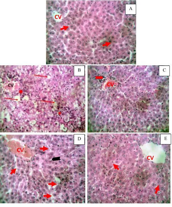
In Figure 3 as shown below, the liver of normal control rats appeared functionally and structurally normal. The hepatocytes showed a well-conserved morphology (1A). The Liver of the Cdcl2-treated group (negative control) showed abnormal changes; there was extensive vacuolation of the hepatocytes with mild infiltration by inflammatory cells (1B).
 Figure 3: Photomicrograph of liver section. (A) Hepatocytes (arrows) are normal with no signs of degenerative lesions/changes. (B) There is extensive vacuolation of the hepatocytes (red arrows) with mild infiltration by inflammatory cells (arrowhead). (C) Hepatocytes are normal; there is mild infiltration of inflammatory cells (arrowhead) around the central vein. (D) Hepatocytes appear normal with mild vacuolations (arrow). (E) The liver section appears normal. Hepatocytes (arrow) and central vein appear normal. CV- central vein [Stain: H and E; ×400].
However, the liver of test group rats (low dose MENS at 300mg/kg) showed mild pericentral vacuolation of hepatocytes with mild infiltration (1C). In the other test group of rats (high dose MENS at 600mg/kg), the liver showed evidence of fatty degeneration (giving a foamy appearance) and vacuolation of some hepatocytes. Furthermore, a photomicrograph of the liver section from CdCl2 + Vitamin C (200mg/kg), showed a normal appearance of hepatocytes (1E).
Let's critically discuss the results
The heart is a muscular organ; made up of cardiac muscles which constantly pump blood through the blood vessels of the circulatory system in the human body and animals; while the kidneys are very effective in the excretion of metabolic wastes. Kidney disease is both a cause and a consequence of cardiovascular disease, hence the term cardiorenal syndrome.
Cardiorenal syndrome is a medical term for disorders involving both the heart and kidneys whereby acute or chronic dysfunction in one organ may induce acute or chronic dysfunction in the other organ. The liver is the main metabolic organ in the body, especially in lipid metabolism and glycogen or glucose metabolism.
Accumulation of toxic substances such as drugs, heavy metals, poisons, etc. can cause significant damage to the histomorphology of vital organs such as the heart muscles, kidney, and liver tissues. Exposure to cadmium could produce serious adverse effects in humans and one of the ways through which this happens is through induction of oxidative stress; whereby it induces early hyperproduction of Reactive Oxygen Species (ROS) that impair antioxidant the defense system leading to oxidative stress in the target organs and tissues.
Always bear this in mind, an imbalance between endogenous antioxidants (produced by the body naturally) and oxidants always results in oxidative stress in cells and tissues. When oxidants are in greater concentration, there is bound to be a change in equilibrium in favour of oxidative stress while if otherwise, cells will not be stressed. We tend to age faster due to the excessive accumulation of these oxidants in the body beyond the scavenging and mopping-up ability of endogenous antioxidants.
In the histopathological analyses, cadmium significantly induced histomorphological changes in the heart fibers, kidney, and liver tissues. However, treatment with low and high-dose methanolic seed extracts of Nigella sativa (MENS) separately, revealed a marked ameliorative effect, thereby protecting the histomorphological architecture of the vital organs studied.
Although this present study was not aimed at evaluating the mechanism through which the seed extract showed ameliorative effects; we however observed that the extract, particularly the low dose, acted in a similar way as the standard drug (Vitamin C), a known antioxidant, which was used (Figures 1-3). Interestingly, we did not observe dose-dependent protection by the seed extract; and these observations once against support the worldwide claim that N.sativa is a potent medicinal plant.
These findings could be a result of the singular or combined actions of one or more of these bioactive phytochemical constituents present in the Nigella sativa. Thymoquinone, an active ingredient of Nigella sativa (black seed), is a constituent of flavonoids.
Several other studies have reported the ameliorative effects- and potential pharmacotherapeutic effects - of crude extracts of N.sativa and thymoquinone separately. N.sativa has been reported to offer protection against cardiotoxic drugs such as cyclosporine A; and against toxic heavy metals such as lead (Pb) and cadmium (Cd).
Crude extracts of the seeds of N. sativa have been evaluated for hepatoprotective activity in Wistar rats against various hepatotoxicants, which are widely known for their ability to induce hepatotoxicity in experimental animals. N. sativa has not only been studied for its protective effects against tissue injuries but it has also been evaluated for antimicrobial activities. Different crude extracts of N. sativa exhibited antimicrobial efficacy against different bacterial strains which comprised either gram-negative or gram-positive bacteria, and these have been reported.
Thymoquinone (TQ) is chemically known as 2-methyl-5-isopropyl-1, 4-benzoquinone. Nemmar et al. reported that TQ showed strong anti-inflammatory effects against diesel exhaust particles-induced cardiopulmonary injury in mice. Due to potent antioxidant and free radical scavenging action, TQ has been shown to normalize the adverse effect of various environmental toxins or xenobiotics causing oxidative damage and organ dysfunctions leading to the pathogenesis of various diseases.
Alkaloids and Phenols, found to be significantly present in the methanol extracts, also act as natural antioxidants, scavenge free radicals and inhibit their production, and stimulate the synthesis of antioxidant enzymes thereby preventing oxidative stress.
Conclusively, the findings of this work show that cadmium induced histomorphological alterations in the heart, kidneys, and liver in the experimental rats; however, methanolic seed extract of Nigella sativa ameliorated the effects in the test groups, although not in a dose-dependent manner. Thus, the result suggests that methanolic seed extract of Nigella sativa (MENS) has anti-cardiotoxic, antinephrotoxic, and hepatoprotective properties and could therefore offer significant amelioration against cadmium-induced organ injuries.
Medicinal plants indeed have huge benefits, we only need to study them critically. Dosage is a very important factor to be considered while administering some of these plant extracts since they contain some phytochemicals that could be toxic at some concentration. That an extract is effective against some disease or pathologic conditions does not expressly means it should be consumed out rightly. It has to undergo toxicity testing to know the right dosage that will not harm cells.
By all means, avoid ingestion of medicinal plants without a regulated dose. Remember, all drugs, are poisons, only one dose makes the safe. Stay safe!
References •Mechanisms of Cell and Tissue Damage •Molecular mechanisms of inflammation and tissue injury after major trauma-is complement the "bad guy"? •Black Cumin (Nigella sativa) and Its Active Constituent, Thymoquinone: An Overview on the Analgesic and Anti-inflammatory Effects •Hepatoprotective effects of Nigella sativa seed extract against acetaminophen-induced oxidative stress •For detailed references and access to the original work on research gate, please click HERE
Return from Original Research: Demonstrating the Protective effects black seed (Nigella sativa) extract has on the heart, kidney and the liver to cyprianj's Web3 Blog

